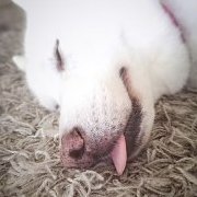
teekay Posted December 12, 2013 Share Posted December 12, 2013 I have posted in the Health forum but didn't get too many replies so thought I would start a thread in general, hoping to get a bit more advice. Jenna has a pink lump on her face. She has had a flat pink spot for a number of weeks but recently it has started to swell. I do not think the diameter has changed but it is getting more pronounced. Went to the vets yesterday morning and she has given Jenna antibiotics for folliculitis. She said to wait a week and if there is no improvement then she will do a fine needle aspiration and or biopsy of it. Here are a couple of pictures of the spot. The first was taken Wednesday, the second today. http://i922.photobucket.com/albums/ad64/tkay67/P1010157_zps6d99b91f.jpg http://i922.photobucket.com/albums/ad64/tkay67/P1010163_zpsbe0af177.jpg It looks to me like it is getting angrier. Not sure whether to wait and see or just ring the vets and get the FNA done sooner. I know the vet will be fine with this and given that I am freaking out I think this may be the best thing to do Just looking for any kind of advice, thoughts, similar stories? Link to comment Share on other sites More sharing options...
hankdog Posted December 12, 2013 Share Posted December 12, 2013 (edited) Thats pretty identical to the two thousand dollar spot Jake had on his face. Started as a tiny nothing then grew to a square centimeter in a week. Two rounds of antibiotics and one of inappropriate surgery and we ended up at the specialist. It was fungal and responded well to a course of Nizoral. He has had a tumour so I freaked out over the whole thing. The specialist just dabbed over it with some Sellotape. Hope yours is the same and nothing to worry about. Edited December 12, 2013 by hankdog Link to comment Share on other sites More sharing options...
pipsqueak Posted December 12, 2013 Share Posted December 12, 2013 (edited) So hard isn't it. Not dismissing hankdog at all... what a terrible expeience. To my very inexperienced eye, it looks like when one of our dogs had a histiocytoma (sp)... which is benign and usually self resolves in ~ 6 weeks... freaked me out because it initially grew rapidly... vet diagnosed with a fine needle biopsy. Edited December 12, 2013 by futuredogtrainer Link to comment Share on other sites More sharing options...
huga Posted December 12, 2013 Share Posted December 12, 2013 You can't really tell until the vet sticks a needle in it. Lola had a lump on her leg earlier this year, she was there for something else, I mentioned it in passing. Vet looked, said it looked like a fatty lump, but would I like him to do a biopsy? I said yes, just to be sure. He was very shocked when he came back in and told me it was a MCT. Link to comment Share on other sites More sharing options...
teekay Posted December 12, 2013 Author Share Posted December 12, 2013 Thanks for the replies. Bloody MCTs, they can differ in appearance so much, buggers Because of where it is, it's going to be impossible to get a good margin on the thing. I think that's why I'm freaking so much. I feel like everyday I am waiting it could be getting that big bigger, deeper etc. Link to comment Share on other sites More sharing options...
White Shepherd mom Posted December 12, 2013 Share Posted December 12, 2013 Casper had something similar looking by his jaw. The vet said it was nothing to worry about and considering his age it was not worth putting him under anaesthetic to remove it. He then found three tumours on his bottom which he said had to come out so the lump on his face was removed at the same time. By the time of the surgery, the lump was so big I thought it would burst at any minute. He certainly does look more handsome without it :) Link to comment Share on other sites More sharing options...
teekay Posted December 12, 2013 Author Share Posted December 12, 2013 Casper had something similar looking by his jaw. The vet said it was nothing to worry about and considering his age it was not worth putting him under anaesthetic to remove it. He then found three tumours on his bottom which he said had to come out so the lump on his face was removed at the same time. By the time of the surgery, the lump was so big I thought it would burst at any minute. He certainly does look more handsome without it :) Did they test it WSM, do you know what it was? Link to comment Share on other sites More sharing options...
Staffyluv Posted December 12, 2013 Share Posted December 12, 2013 After my experience with MCTs and Ollie dog - I would never settle for anything less than an immediate FNA on every lump found.. MCTs can look like a pea under the skin, a wart (varying in colour) and even a fatty lump (which is usually suggested as a lipoma).. MCTs can get out of hand so quickly, the wait and see approach just doesn't appeal to me at all.. Link to comment Share on other sites More sharing options...
Guest Maeby Fünke Posted December 12, 2013 Share Posted December 12, 2013 I would get the lump tested ASAP. I worry that if I leave a lump too long the cancer (if it is cancerous) will become a higher grade. Having it tested early gives me time to think about how I'm going to deal with it. There's nothing worse than feeling panicked and upset and having to make a decision about cancer treatment. MCT's come in all shapes and sizes. You never know until you've had a biopsy, or a fine needle aspirate at the very least. Hoping it's nothing to worry about :) Link to comment Share on other sites More sharing options...
teekay Posted December 12, 2013 Author Share Posted December 12, 2013 Thanks all. I've just called the vets. Jenna is going in tomorrow for the FNA. I need to know one way or the other. Link to comment Share on other sites More sharing options...
Guest Maeby Fünke Posted December 12, 2013 Share Posted December 12, 2013 (edited) Don't worry about the margins. If it is an MCT and it's a LOW grade one it won't need clean margins. That is now considered out-dated thinking by oncologists. eta My Pug has had MCT's removed from his nose roll, mouth, ear and lower leg without clean margins. Sorry, I meant LOW grade! Edited December 12, 2013 by Maeby Fünke Link to comment Share on other sites More sharing options...
Guest Maeby Fünke Posted December 12, 2013 Share Posted December 12, 2013 Thanks all. I've just called the vets. Jenna is going in tomorrow for the FNA. I need to know one way or the other. Sorry, I didn't see your post. I hope it all goes well :) Link to comment Share on other sites More sharing options...
~Anne~ Posted December 12, 2013 Share Posted December 12, 2013 The FNA cannot tell you what it is. It can rule out some things though. For example, if the lump has fatty cells then it is usually a lipoma I believe. If the cells are more fibrous it could be several types of tumour or none. That's my understanding anyway. Boof has yet another on his neck. It looks innocuous but with his history we can't be sure. This one is like a small wart. He's had one before that looked like a wart and it was an MCT. He's had MCTs that look like fatty lumps and he's had one way under the dermis. They're insidious little things and can take all different shapes and appearances. Link to comment Share on other sites More sharing options...
Guest Maeby Fünke Posted December 12, 2013 Share Posted December 12, 2013 (edited) Sorry, that didn't come out right... A fine needle aspirate can diagnose mast cell tumours, but a biopsy is better than a fine needle aspirate because it will tell you what grade the cancer is. eta Sorry, I don't mean to be pedantic, I'm just trying to be clear. I'm wary of giving people false information about such a serious subject. Edited December 12, 2013 by Maeby Fünke Link to comment Share on other sites More sharing options...
Guest Maeby Fünke Posted December 12, 2013 Share Posted December 12, 2013 (edited) The FNA cannot tell you what it is. It can rule out some things though. For example, if the lump has fatty cells then it is usually a lipoma I believe. If the cells are more fibrous it could be several types of tumour or none. That's my understanding anyway. Boof has yet another on his neck. It looks innocuous but with his history we can't be sure. This one is like a small wart. He's had one before that looked like a wart and it was an MCT. He's had MCTs that look like fatty lumps and he's had one way under the dermis. They're insidious little things and can take all different shapes and appearances. Oh no, Anne. I hope it's nothing My Pug has another one on his chin. I just noticed it yesterday and I'm taking him in on Monday. This will be his 15th tumour! Edited December 12, 2013 by Maeby Fünke Link to comment Share on other sites More sharing options...
hankdog Posted December 12, 2013 Share Posted December 12, 2013 On Maeby I hope he's ok. So worrisome when they have health problems. Link to comment Share on other sites More sharing options...
Guest Maeby Fünke Posted December 12, 2013 Share Posted December 12, 2013 On Maeby I hope he's ok. So worrisome when they have health problems. Thanks :) It is a bit concerning, but I'm going to be really tough with myself and not worry about it until I know what it is. Link to comment Share on other sites More sharing options...
Guest Clover Posted December 12, 2013 Share Posted December 12, 2013 Good luck for tomorrow Teekay. Everything crossed for you and Jenna. I don't leave the vets without having at least a fna done when it comes to lumps. My little scruffer had a lump checked the other day and it was just fatty at this stage but we need to keep an eye on it. Link to comment Share on other sites More sharing options...
teekay Posted December 13, 2013 Author Share Posted December 13, 2013 Hope your pugs are ok Anne and Maeby. Bloody lumps and bumps, I hate them Vet just called and she did a couple of FNAs and is only seeing inflammatory cells so she is fairly confident her diagnosis of an infection is correct. She did say she cannt be absolutely certain without a biopsy what it is, but she did say she could rule out an MCT. She did give me the option of doing a biopsy while Jenna is still sedated which would cost about $700 Given she is confident it is not an MCT I decided to give the antibiotics longer to do their job and if we are not seeing an improvement in a couple of weeks we will biopsy it then. Feeling a lot happier and looking forward to picking her up later. :) Thanks all of you Link to comment Share on other sites More sharing options...
persephone Posted December 13, 2013 Share Posted December 13, 2013 :) The AB's will kick in ..skin often seems to take ages - just look at how long human pimples can hang around!. perhaps dab some colloidal silver on it a few times a day , too ..it gets absorbed and helps fight bacteria on/just under the surface . Link to comment Share on other sites More sharing options...
Recommended Posts
Create an account or sign in to comment
You need to be a member in order to leave a comment
Create an account
Sign up for a new account in our community. It's easy!
Register a new accountSign in
Already have an account? Sign in here.
Sign In Now